Neurology and Brain Health
White Matter Spots on Brain MRI: What They Mean and What to Do Next
A report describing white matter hyperintensities can sound alarming. These MRI findings become more common with age, but their cause and importance depend on their pattern, burden, symptoms, and vascular risk factors.
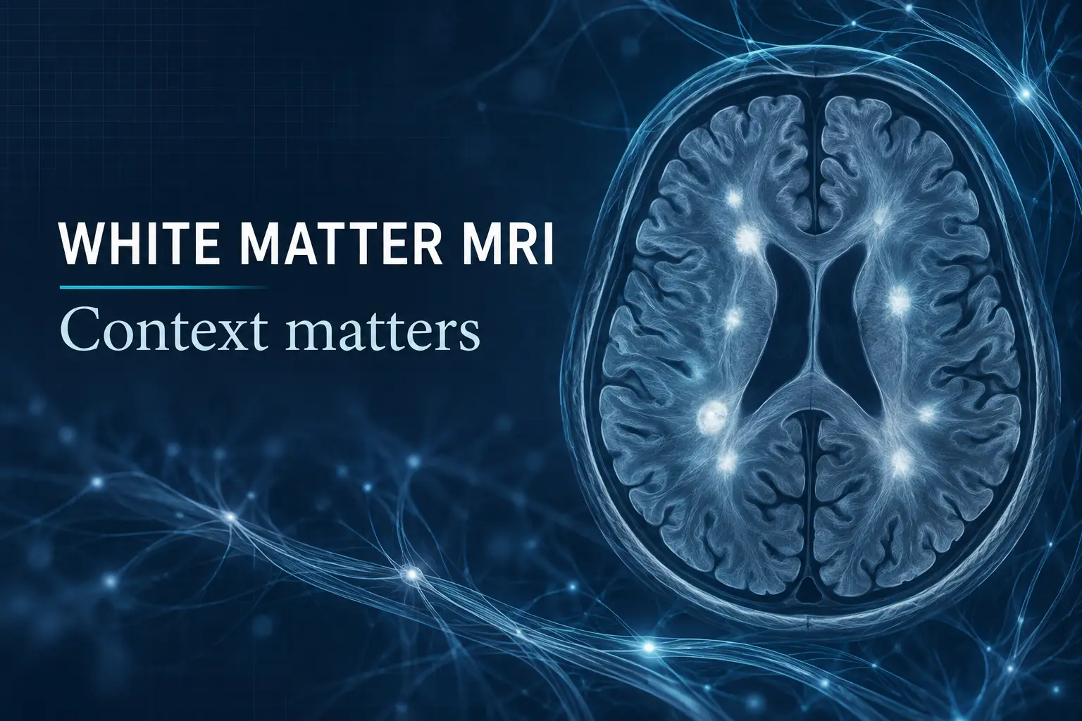
A brain MRI report may use phrases such as “white matter hyperintensities,” “nonspecific white matter changes,” “chronic microvascular ischemic disease,” or “leukoaraiosis.” Patients often read these words and wonder whether they have had a stroke, are developing multiple sclerosis, or will develop dementia.
The answer is rarely contained in one phrase. White matter spots are common, especially with increasing age, but they are not all interchangeable. Their significance depends on why the MRI was ordered, the person’s age and symptoms, the number and location of lesions, whether they are changing, and what the radiologist thinks the pattern most likely represents.
What are white matter hyperintensities?
White matter contains bundles of nerve fibers that connect different brain regions. On certain MRI sequences, particularly T2 and FLAIR images, some areas can appear brighter than the surrounding tissue. Radiologists call these areas white matter hyperintensities, or WMH.
The term describes an imaging appearance, not a single disease. These bright areas reflect increased water content in tissue and can arise through different mechanisms. In adults over 50, many are attributed to changes in the brain’s small blood vessels. Other possibilities include migraine-associated changes, inflammation, demyelinating disease, prior injury, infection, genetic small-vessel disorders, and less common conditions.
The radiologist’s description matters. Small, scattered, symmetrical spots in an older adult with hypertension are interpreted differently from lesions in the corpus callosum, brainstem, or spinal cord in a younger person with new neurologic symptoms.
Are white matter spots a normal part of aging?
They are common, but “common” does not mean “meaningless.” A 2023 consensus statement reported that WMH are present in roughly half of people in their 40s and in as many as 95% of people by age 90. The burden varies widely.
Small, stable lesions may never cause noticeable symptoms. More extensive or confluent WMH, however, are associated in population studies with higher risks of stroke, cognitive decline, gait difficulty, depression, disability, and death. Association is not destiny. These studies describe group-level risk and cannot predict exactly what will happen to one person.
WMH also do not prove Alzheimer’s disease. A 2021 meta-analysis of 36 prospective studies found that a greater WMH burden was associated with increased risk of cognitive impairment and dementia, including vascular dementia. That does not establish that the spots alone caused dementia, nor does it mean that everyone with WMH will develop it.
The most useful response is neither panic nor dismissal. The finding can be treated as a prompt to assess brain and cardiovascular health.
Which risk factors matter most?
Age and high blood pressure are among the strongest recognized factors associated with vascular WMH. Diabetes, smoking, abnormal cholesterol, and other cardiovascular risks may also contribute. Obstructive sleep apnea has been associated with greater cerebral small-vessel disease in observational research, although it has not been proven that apnea treatment reverses existing WMH.
Migraine deserves nuance. People with migraine can have WMH, often without the same vascular implications seen in someone with multiple cardiovascular risk factors. The clinical history and lesion pattern are essential before assigning meaning.
A clinician evaluating incidental WMH may review:
- Blood pressure, including whether treatment is reaching an appropriate goal
- Blood glucose or hemoglobin A1c
- Cholesterol and overall cardiovascular risk
- Smoking or nicotine exposure
- Physical activity, diet, weight, and medication history
- Prior episodes of weakness, numbness, speech trouble, vision loss, imbalance, or confusion
- Symptoms suggesting a sleep disorder when clinically appropriate
The 2024 American Heart Association and American Stroke Association guideline emphasizes controlling established vascular risks, especially hypertension, to prevent a first stroke. The appropriate targets and medications must be individualized.
Should you start aspirin because of white matter changes?
Not automatically. The 2023 consensus statement specifically advises against starting an antiplatelet drug, such as aspirin, or an anticoagulant solely because incidental WMH were found, unless another medical indication exists.
Aspirin can reduce clot-related events in selected patients, but it can also cause bleeding. The decision depends on a person’s complete cardiovascular and cerebrovascular history, not the presence of nonspecific white matter spots alone. Do not start or stop aspirin based only on an MRI phrase.
Does every patient need a neurologist or another MRI?
No single schedule fits everyone. Typical mild vascular-appearing WMH may be managed initially by the clinician who ordered the scan or a primary care physician, with attention to risk-factor control.
A neurology evaluation becomes more important when:
- Symptoms are new, progressive, recurrent, or unexplained
- The person is younger than expected for the amount of disease
- The MRI distribution is atypical for ordinary small-vessel change
- The report raises demyelination, inflammation, vasculitis, infection, genetic disease, or another specific diagnosis
- There are cognitive, gait, balance, bladder, seizure, or other neurologic concerns
- Prior images show unexpectedly rapid progression
Repeat imaging should answer a clinical question. It may be appropriate when the pattern is uncertain, symptoms change, or a disease requiring surveillance is suspected. Routine serial MRI is not automatically necessary for every stable incidental finding.
What can you do now?
The most practical next step is to review the actual report and images in clinical context. Ask what pattern the radiologist described, whether the burden is mild or more extensive, and whether the lesions are typical for age and risk factors.
If the findings are believed to be vascular, focus on measures with established health benefits:
- Measure and appropriately treat high blood pressure
- Address diabetes and abnormal cholesterol
- Avoid smoking
- Exercise regularly within medical limitations
- Follow a heart-healthy eating pattern
- Treat relevant sleep disorders and maintain consistent, adequate sleep
- Take prescribed medication consistently
These measures support overall cardiovascular and brain health. It is not yet proven that any one lifestyle intervention will erase existing WMH or guarantee that they will not progress.
When is it an emergency?
Chronic white matter changes on a report are not usually an emergency by themselves. New neurologic symptoms are different.
Call 911 for sudden facial droop, one-sided weakness or numbness, speech difficulty, vision loss, severe imbalance, confusion, or an abrupt severe headache. A transient ischemic attack can resolve quickly but still requires emergency evaluation. There is no reliable way at symptom onset to know whether an event is a TIA or a completed stroke.
Frequently Asked Questions About White Matter Spots
Do white matter spots mean I have dementia?
No. White matter hyperintensities are associated with cognitive risk at the population level, but they do not prove Alzheimer’s disease or predict one person’s future. Age, lesion burden, pattern, symptoms, and vascular risks all matter.
Are white matter spots the same as multiple sclerosis?
Not necessarily. Many white matter spots reflect small-vessel change, migraine, or other causes. Lesion location, shape, clinical history, examination, and sometimes follow-up imaging help distinguish an MS-like pattern from more common explanations.
Can white matter spots disappear?
Established lesions often remain visible. The more useful goal is to identify the cause, control modifiable risks, and decide whether symptoms or an atypical MRI pattern require further evaluation.
The bottom line
White matter spots are an MRI finding, not a diagnosis. Many are common and mild, but a larger or atypical burden can provide useful information about vascular or neurologic health. The right response is to interpret the pattern, review symptoms, identify modifiable risks, and avoid treatments that are not otherwise indicated.
Brainology’s neurology and brain-health team can evaluate white matter changes in the context of symptoms, neurologic examination, vascular risks, sleep health, and the complete MRI pattern. The goal is a clear plan based on the individual scan and patient, not fear created by a single line in a report.
This article is for general educational purposes and does not provide a diagnosis or individualized treatment plan. New stroke-like symptoms require emergency care.
References
- Ottavi TP, et al. Consensus statement for the management of incidentally found brain white matter hyperintensities in general medical practice. Medical Journal of Australia. 2023.
- Bushnell C, et al. 2024 Guideline for the Primary Prevention of Stroke: A Guideline From the American Heart Association/American Stroke Association. Stroke. 2024.
- Smith EE, et al. Prevention of Stroke in Patients With Silent Cerebrovascular Disease: A Scientific Statement From the American Heart Association/American Stroke Association. Stroke. 2017.
- Debette S, et al. Clinical Significance of Magnetic Resonance Imaging Markers of Vascular Brain Injury: A Systematic Review and Meta-analysis. JAMA Neurology. 2019.
- Hu HY, et al. White matter hyperintensities and risks of cognitive impairment and dementia: A systematic review and meta-analysis of 36 prospective studies. Neuroscience & Biobehavioral Reviews. 2021.
- Brown R, et al. Rate of, and risk factors for, white matter hyperintensity growth: a systematic review and meta-analysis. Journal of Neurology, Neurosurgery & Psychiatry. 2021.
- Centers for Disease Control and Prevention. About Stroke. Updated October 24, 2024.