Neurology and Brain Health
Walking, Memory, and Bladder Changes: When to Consider Normal Pressure Hydrocephalus
Normal pressure hydrocephalus can resemble other neurologic conditions. The pattern of gait change, cognitive symptoms, bladder problems, imaging, and response to cerebrospinal fluid drainage helps guide diagnosis.
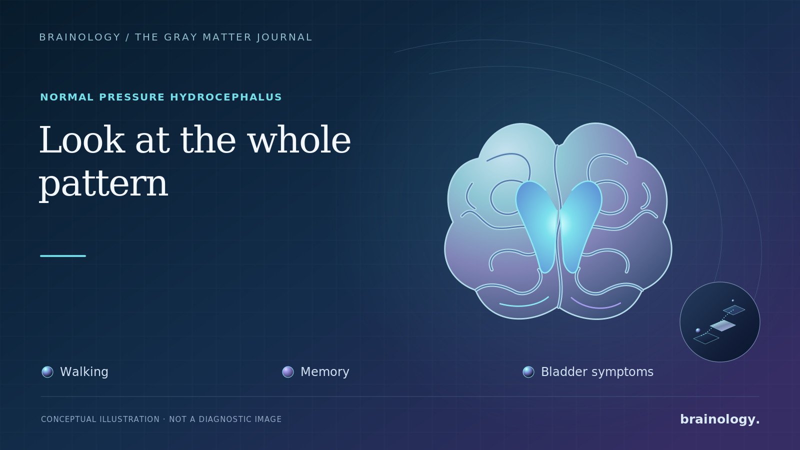
An older adult begins taking shorter steps. Turning becomes awkward, and getting started feels as if the feet are stuck to the floor. Months later, family members notice slower thinking or difficulty organizing everyday tasks. Urinary urgency may appear too.
Each symptom has many possible causes. Together, however, they can raise the question of normal pressure hydrocephalus, or NPH.
NPH deserves attention because some appropriately selected patients improve after surgery. It also deserves caution. The symptoms are not unique to NPH, an enlarged fluid space on a brain scan does not establish the diagnosis, and treatment carries meaningful risks. Evaluation is a process, not a single test.
What Is Normal Pressure Hydrocephalus?
Cerebrospinal fluid, or CSF, surrounds the brain and spinal cord and circulates through spaces inside the brain called ventricles. In NPH, the ventricles become enlarged in association with a characteristic clinical syndrome.
The term “normal pressure” can be confusing. A pressure measurement obtained during a lumbar puncture may fall within the usual range, but that does not mean CSF circulation and pressure dynamics are normal at all times.
Some cases follow a known event, such as bleeding around the brain, meningitis, head trauma, or another condition that disrupts CSF flow. When no clear cause is found, the condition is called idiopathic normal pressure hydrocephalus, or iNPH. It primarily affects older adults.
The Classic Triad Is a Clue, Not a Checklist
NPH is commonly taught as a triad of gait impairment, cognitive decline, and urinary incontinence. Real patients do not always arrive with all three symptoms at the same time.
Gait impairment is often the most prominent clue. A person may walk slowly with short steps and a wider base. Starting, turning, or navigating a narrow space can be especially difficult. The feet may appear “magnetic,” although that description is not present in every patient. Balance problems and falls can follow.
Cognitive changes may involve processing speed and executive function. Families might notice reduced initiative, slowed responses, difficulty planning, or trouble managing several steps of a task. Memory can be affected, but the pattern may differ from the early memory-predominant presentation often associated with Alzheimer’s disease.
Bladder symptoms may begin with urgency and frequency. Incontinence can develop later. Urinary symptoms are common in older adults and may result from prostate disease, urinary tract problems, medications, mobility limitations, diabetes, or other neurologic disorders. They become more informative when they develop alongside the right gait and cognitive pattern.
The absence of one part of the triad does not automatically rule out NPH. Conversely, having all three does not prove it.
Why NPH Can Be Difficult to Diagnose
The same symptoms can occur with several common conditions. Parkinson’s disease and related disorders can slow gait and impair balance. Arthritis, peripheral neuropathy, spinal stenosis, cervical spinal cord compression, vestibular disease, sedating medications, and muscle weakness can also affect walking. Alzheimer’s disease, vascular cognitive impairment, depression, sleep disorders, medication effects, and metabolic problems can affect cognition.
More than one condition may be present. An individual can have enlarged ventricles and Alzheimer’s pathology, vascular brain disease, or a separate gait disorder. Treating one contributor may help without resolving every symptom.
That is why a careful assessment usually includes the history and timeline, neurologic examination, medication review, objective gait testing, cognitive evaluation, and brain imaging. Depending on the findings, clinicians may also evaluate the spine, peripheral nerves, sleep, laboratory abnormalities, or urologic causes.
NPH typically causes a gradual or progressive pattern. Sudden facial droop, one-sided weakness, speech difficulty, severe headache, abrupt confusion, or an acute inability to walk should be treated as a possible emergency, not assumed to be NPH.
What Brain Imaging Can Show
CT or MRI can demonstrate enlarged ventricles. Clinicians also look at the shape of the ventricles and the distribution of fluid spaces around the brain. A pattern called disproportionately enlarged subarachnoid-space hydrocephalus, or DESH, can support suspicion for iNPH.
Imaging must be interpreted with the clinical picture. Ventricles can enlarge when brain tissue has been lost through aging, neurodegeneration, stroke, or other disease. This is sometimes called enlargement “ex vacuo.” A scan report that mentions ventriculomegaly does not, by itself, establish NPH or indicate that a shunt is needed.
Likewise, no single imaging feature perfectly predicts who will improve with surgery. The decision rests on converging evidence.
Why Clinicians May Recommend a Tap Test
During a CSF tap test, a clinician performs a lumbar puncture and removes a measured amount of cerebrospinal fluid. Walking is assessed before and after drainage, ideally with standardized measures such as gait speed, step length, turning, or a timed walking test. Some centers also reassess cognition and bladder symptoms.
Improvement after CSF removal strengthens the likelihood that shunting will help. The timing of improvement varies, so testing may be repeated over several hours or the following day. In selected cases, clinicians use extended lumbar drainage or specialized CSF flow and pressure testing.
A negative tap test does not necessarily close the case. Current guidelines recognize that some patients who do not show a clear short-term response still improve after shunting. Measurement technique, fatigue, pain, delayed response, other illnesses, and the outcome chosen can affect the result. Further evaluation should be individualized rather than based on one yes-or-no finding.
What Does Shunt Surgery Actually Improve?
The usual treatment is surgical placement of a shunt that diverts CSF from a brain ventricle to another part of the body, most often the abdomen. Programmable valves allow clinicians to adjust drainage without another operation.
The strongest recent evidence comes from the 2025 PENS trial, a multicenter, double-blind, placebo-controlled study of 99 participants. All had findings consistent with iNPH and had improved gait velocity after temporary CSF drainage. Three months after surgery, participants whose shunts were open had greater improvement in gait speed and gait-and-balance scores than those whose valves were initially set to provide essentially no drainage.
The trial did not find significant between-group improvement at three months on its screening measures of cognition or overactive bladder symptoms. Longer follow-up is important, and a brief cognitive screening test may not capture every meaningful change. Still, the result argues against promising that shunting will reverse every part of the syndrome.
Surgery also has risks. In the PENS trial, subdural bleeding and positional headaches occurred more often in the open-shunt group, while falls were more frequent in the placebo group. Potential complications in practice include infection, bleeding, excessive or inadequate drainage, obstruction, and the need for valve adjustments or additional surgery.
Earlier AAN guidance concluded that shunting was possibly effective, especially for gait, but emphasized weighing expected benefit against adverse events. The newer randomized evidence provides stronger support for gait improvement in carefully selected patients. It does not make patient selection or risk discussion less important.
When Should a Family Ask About NPH?
Consider discussing NPH with a clinician when an older adult develops a progressive, unexplained gait disorder, especially when slower thinking or new urinary urgency accompanies it. Useful information to bring includes:
- When each symptom began and how it has changed
- A complete medication list
- Details about falls and mobility aids
- Prior brain scans and their reports
- Brief videos of walking at home, if safe to obtain
- Observations from someone who knows the person well
Ask what alternative diagnoses are being considered, whether the ventricular enlargement matches the clinical pattern, how gait will be measured before and after CSF drainage, and which symptoms are realistically expected to improve.
NPH is neither a diagnosis to overlook nor an explanation to force onto every combination of walking and memory problems. The best evaluation integrates the person’s symptoms, examination, imaging, response to CSF drainage, other medical conditions, goals, and surgical risk.
Brainology can provide neurologic evaluation of progressive gait and cognitive symptoms, coordinate appropriate imaging and testing, and help patients and families understand when specialty hydrocephalus or neurosurgical assessment may be appropriate.
This article is for educational purposes and does not replace individualized medical care. Seek emergency evaluation for sudden neurologic symptoms. Decisions about lumbar puncture or shunt surgery require assessment by qualified clinicians.
References
- National Institute of Neurological Disorders and Stroke. Hydrocephalus. Updated May 19, 2026.
- Nakajima M, Yamada S, Miyajima M, et al. Guidelines for Management of Idiopathic Normal Pressure Hydrocephalus, Third Edition. Neurologia Medico-Chirurgica. 2021;61(2):63-97. doi:10.2176/nmc.st.2020-0292.
- Luciano MG, Williams MA, Hamilton MG, et al. A Randomized Trial of Shunting for Idiopathic Normal-Pressure Hydrocephalus. New England Journal of Medicine. 2025;393(22):2198-2209. doi:10.1056/NEJMoa2503109.
- Halperin JJ, Kurlan R, Schwalb JM, Cusimano MD, Gronseth G, Gloss D. Practice guideline: Idiopathic normal pressure hydrocephalus: Response to shunting and predictors of response. Neurology. 2015;85(23):2063-2071. doi:10.1212/WNL.0000000000002193.
- Pearce RKB, Gontsarova A, Richardson D, et al. Shunting for idiopathic normal pressure hydrocephalus. Cochrane Database of Systematic Reviews. 2024;8:CD014923. doi:10.1002/14651858.CD014923.pub2.
- Carswell C. Idiopathic normal pressure hydrocephalus: historical context and a contemporary guide. Practical Neurology. 2023;23(1):15-22. doi:10.1136/practneurol-2021-003291.
- Williams MA, Nagel SJ, Golomb J, et al. Safety and effectiveness of the assessment and treatment of idiopathic normal pressure hydrocephalus in the Adult Hydrocephalus Clinical Research Network. Journal of Neurosurgery. 2022;137(5):1289-1301. doi:10.3171/2022.1.JNS212782.