Neurology and Brain Health
Can You Have Epilepsy With a Normal EEG?
A normal routine EEG does not rule out epilepsy. EEG is one part of a clinical evaluation that may also include a detailed event history, witness video, brain imaging, and longer monitoring.
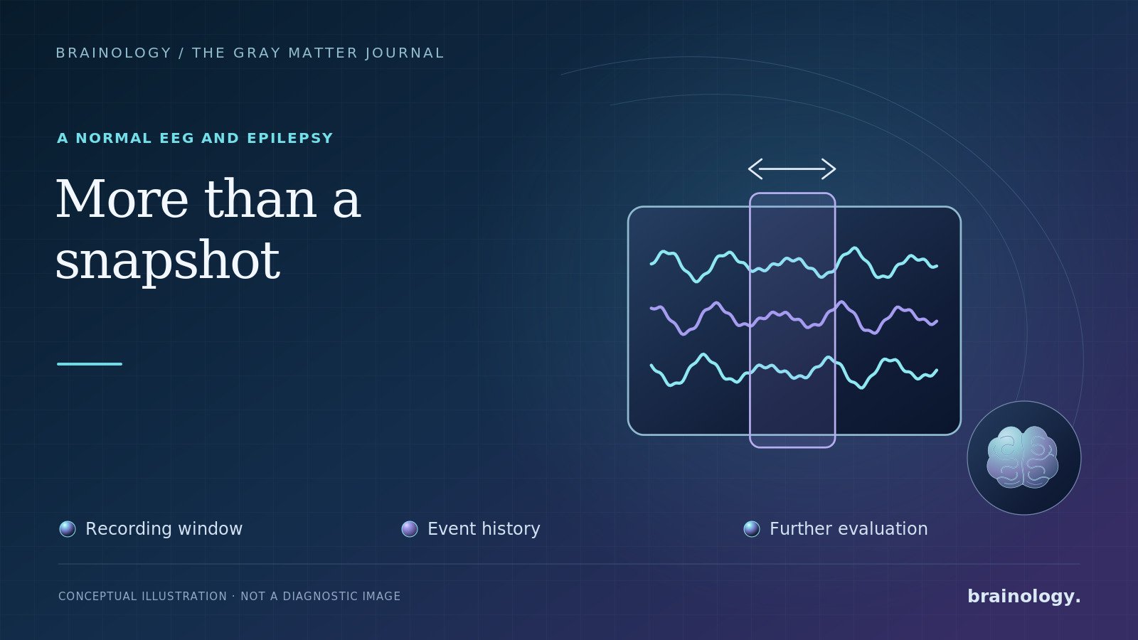
Yes. A person can have epilepsy even when a routine electroencephalogram, or EEG, is normal.
That answer can feel confusing. EEG is often described as a test for seizures, so patients may expect a simple positive or negative result. In practice, an EEG is a sample of the brain's electrical activity during the recording. Epileptic abnormalities may be intermittent, and a short study may not capture them.
A normal result can be reassuring in some respects, but it does not erase a convincing event history. An abnormal result can provide useful evidence, but it does not establish epilepsy by itself. Diagnosis depends on how the episode happened, what witnesses observed, the EEG findings, imaging, and other possible explanations.
What a Routine EEG Actually Records
Small electrodes placed on the scalp measure electrical activity generated by groups of brain cells. A routine study usually records wakefulness and may capture drowsiness or sleep. Laboratories may also use deep breathing and flashing lights to reveal abnormalities that are not visible during quiet wakefulness.
EEG can help a neurologist:
- Identify interictal epileptiform discharges, which are abnormal patterns seen between seizures
- Support the diagnosis of epilepsy when the clinical history is compatible
- Help classify whether epilepsy appears focal, generalized, or part of a particular syndrome
- Estimate the risk of another seizure after a first unprovoked event
- Guide the choice of antiseizure medication in selected cases
Most routine EEGs do not record an actual seizure. The test is often looking for brief abnormalities between events.
Why the EEG Can Be Normal
Epileptiform discharges may occur only occasionally. They can also become more visible during sleep, after sleep deprivation, or at certain times of day. A focal abnormality arising deep in the brain may not create a clear signal at the scalp. Medications and the timing of the study can also affect what appears.
In a 2023 prospective study of 100 adults evaluated after a first unprovoked seizure, 24-hour ambulatory EEG detected epileptiform discharges or seizures with greater sensitivity than either the first or second routine EEG. Sensitivity was 72% for ambulatory EEG, compared with 11% for the first routine EEG and 22% for the second. Those exact numbers should not be applied to every clinic or patient, but they illustrate why a longer recording may find abnormalities missed during a shorter sample.
International EEG standards also note that sensitivity increases with repeated recordings and that a sleep EEG can add diagnostic value when an initial routine study is unrevealing.
A Normal EEG Does Not Confirm That an Event Was Nonepileptic
A normal EEG cannot prove that a spell was fainting, migraine, a sleep disorder, a functional seizure, or another nonepileptic event. It only means that the recording did not show the abnormalities being sought during that period.
Likewise, not every collapse, stare, shaking episode, or period of confusion is epilepsy. Possible alternatives include syncope caused by a blood-pressure or heart-rhythm problem, low blood sugar, medication or substance effects, migraine, sleep disorders, and functional seizures. Some people can have more than one condition.
The event description is therefore crucial. Details such as warning symptoms, head or eye position, responsiveness, movements, color change, injuries, duration, and recovery may be more informative than a brief recollection from the person who lost awareness. A safe smartphone video recorded by a witness can sometimes help, but no one should delay first aid or place themselves in danger to obtain one.
An Abnormal EEG Is Not a Diagnosis by Itself
The opposite misunderstanding also occurs. Sharp-looking waves, artifacts, and normal variants can be mistaken for epileptiform activity. Rare epileptiform abnormalities can even appear in people without a known seizure history.
For that reason, an EEG should be interpreted by a qualified clinician in the context of the patient's symptoms. Treating an EEG tracing rather than the person can lead to an incorrect diagnosis, unnecessary medication, driving restrictions, anxiety, and missed alternative causes.
What May Come Next After a Normal Routine EEG
The next step depends on how strongly the history suggests a seizure and how urgently an answer is needed. Options may include:
Repeat or sleep-deprived EEG
A repeat study gives another opportunity to capture an interictal abnormality. Sleep and the transition between sleep and wakefulness can activate epileptiform discharges. Sleep deprivation should be used only as instructed because it can increase seizure risk and make driving unsafe.
Ambulatory EEG
Ambulatory monitoring records brain activity for a day or longer while the patient follows much of a normal routine. It increases sampling time and may capture sleep or a typical event. Many systems also record video, although home video coverage can be incomplete.
Inpatient video EEG
Continuous video EEG in an epilepsy monitoring unit can match behavior on video with the electrical recording. It is especially useful when recurrent events remain unexplained, event classification will change treatment, or functional seizures and epilepsy must be distinguished. Not every patient needs inpatient monitoring.
Brain MRI with an epilepsy protocol
EEG evaluates electrical function. MRI evaluates structure. A dedicated epilepsy-protocol MRI can identify lesions that a routine CT or general MRI may miss. Imaging findings must still be matched with the event history and EEG rather than assumed to be the cause.
What a First Seizure Means for Future Risk
One unprovoked seizure does not automatically mean a person has epilepsy. The American Academy of Neurology and American Epilepsy Society estimate that the risk of another seizure is highest during the first two years, with an overall range of 21% to 45% in the evidence they reviewed.
Factors associated with higher recurrence risk include a prior brain injury, epileptiform abnormalities on EEG, a significant brain-imaging abnormality, and a seizure during sleep. These factors help guide an individualized discussion about whether to begin medication after a first event. Immediate treatment can reduce short-term recurrence but has not been shown to improve the long-term chance of sustained remission.
Safety Matters While the Evaluation Is Ongoing
After a possible seizure, follow driving restrictions and obtain medical clearance before returning to the road. Laws differ by location. Until the risk is clearer, avoid swimming alone, baths without supervision, unprotected heights, open flames, and operating dangerous machinery.
During a convulsive seizure, protect the person from injury, place them on their side when possible, and time the event. Do not restrain them or put anything in their mouth. Call 911 for a first seizure, an event lasting longer than five minutes, repeated seizures without recovery, trouble breathing or waking, a serious injury, a seizure in water, or a seizure during pregnancy.
The Bottom Line
A normal routine EEG is not the same as "no epilepsy." It is one result from a limited recording window. The most useful interpretation combines the EEG with a careful history, witness information, neurologic examination, appropriate imaging, and, when needed, longer video EEG monitoring.
Brainology can help evaluate unexplained spells, review prior EEG or imaging results, and determine whether additional testing is appropriate. The goal is not simply to find an abnormal waveform. It is to reach the most accurate diagnosis while avoiding unnecessary treatment.
This draft is for educational purposes and clinical and editorial review. It is not a diagnosis or a substitute for emergency care or individualized medical advice.
References
- Krumholz A, Wiebe S, Gronseth GS, et al. Practice Parameter: Evaluating an Apparent Unprovoked First Seizure in Adults. Neurology. 2007;69(21):1996-2007. PubMed
- Krumholz A, Wiebe S, Gronseth GS, et al. Evidence-Based Guideline: Management of an Unprovoked First Seizure in Adults. Neurology. 2015;84(16):1705-1713. PubMed
- Hernandez-Ronquillo L, Thorpe L, Feng C, et al. Diagnostic Accuracy of Ambulatory EEG vs Routine EEG in Patients With First Single Unprovoked Seizure. Neurology: Clinical Practice. 2023;13(3):e200160. PubMed
- Peltola ME, Leitinger M, Halford JJ, et al. Routine and Sleep EEG: Minimum Recording Standards of the International Federation of Clinical Neurophysiology and the International League Against Epilepsy. Clinical Neurophysiology. 2023;147:108-120. PubMed
- Fisher RS, Acevedo C, Arzimanoglou A, et al. ILAE Official Report: A Practical Clinical Definition of Epilepsy. Epilepsia. 2014;55(4):475-482. ILAE
- Bernasconi A, Cendes F, Theodore WH, et al. Recommendations for the Use of Structural Magnetic Resonance Imaging in the Care of Patients With Epilepsy. Epilepsia. 2019;60(6):1054-1068. PubMed
- Centers for Disease Control and Prevention. First Aid for Seizures. Updated May 15, 2024. CDC